3D-Printed Phantom Models for Medical Imaging
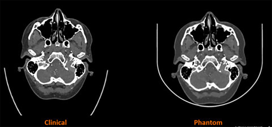
Stratasys and Siemens Healthineers partnered to develop a technology that translates information from scanned patient images (CT) into 3D-printed phantom models with specific material characteristics that emulate the radiopacity of human anatomy.
Medical imaging has used phantoms for computed tomography (CT) for a while since they serve as specialized tools for assessing and verifying the functionality of CT scanners. These phantoms are tailored to replicate specific attributes of the human body and facilitate the evaluation of crucial parameters such as radiation dose and image quality. This process supports calibration efforts and ensures the consistent performance of the scanner.
It is more than a 3D model, radiopaque accuracy is needed
In collaboration, Stratasys and Siemens Healthineers integrate Stratasys’ PolyJet™ technology, its proprietary RadioMatrix™ technology, and Siemens Healthineers’ advanced algorithm. This combination aims to translate scanned patient images into specific material characteristics, achieving the radiopacity of human anatomy.
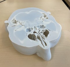
3D-printed phantom head slice. Courtesy of Stratasys.
The solution will allow for tailored phantom manufacturing and the creation of ultra-realistic human anatomy characteristics. A complete radiographic accuracy of patient-specific pathology not previously possible.
This joint project will transform how the medical field utilizes phantoms. In certain cases, even enable device manufacturers and academic facilities to replace human cadavers with 3D-printed structures.
“The current limitations of imaging phantoms have been a longstanding challenge for the radiology community,” said Erez Ben Zvi, Vice President of Medical at Stratasys. “This partnership with Siemens Healthineers will enable us to jointly explore the vast possibilities of our radiopaque materials and 3D printing technologies to overcome these barriers.”
From smaller parts to a human torso
This project will create important research data, giving valuable insights to improve algorithms for computer tomography (CT) systems. It will also contribute to the development of materials, and open up possibilities for exploring new applications.
The project begins by producing 3D-printed models for smaller parts such as the head and neck. As the research advances, progressively larger and more detailed body parts will take shape. The goal is to initially 3D print a heart model and eventually create a detailed entire human torso.

Juliana Montoya is Director of Content for Plastics Engineering. A mechanical engineer with an MSc in materials engineering, she has experience as a sustainability and packaging consultant focused on ecodesign and recycling. Her work centers on technical content for the plastics industry, connecting polymer innovation, manufacturing trends, and sustainability strategy for industry audiences.