Wearable Ultrasound Patch Scans for Breast Cancer Tumors
MIT researchers say the device changes the form factor of the ultrasound technology
MIT researchers have developed a wearable ultrasound device that could help patients detect breast cancer tumors sooner, thereby improving their survival chances. The device, which can be incorporated in a bra, is designed to allow more frequent monitoring of patients at high risk for the disease.
The survival rate for breast cancer detected in its earliest stages is nearly 100 percent, with that dropping to about 25 percent for tumors detected at later stages. Breast tumors that develop in between regularly scheduled mammograms — known as interval cancers — account for 20 to 30 percent of all breast cancer cases, and these tumors tend to be more aggressive than those found during routine scans.

Dr. Canan Dagdeviren
Canan Dagdeviren, an associate professor in MIT’s Media Lab said she was inspired to pursue this project after her aunt, despite having regular cancer screenings, was diagnosed with late-stage breast cancer at age 49 and passed away six months later.
Dagdeviren, a Fulbright Scholar and a native of Turkey, received her Ph.D. in Material Science and Engineering from the University of Illinois Urbana-Champaign in 2014. She is the senior author of a recent study on the project, titled “Conformable ultrasound breast patch for deep tissue scanning and imaging.” The journal Science Advances published the paper on July 28. It lists MIT graduate student Wenya Du, Research Scientist Lin Zhang, Emma Suh ’23, and Dabin Lin, a professor at Xi’an Technological University, as the lead authors. (Here is a short interview with Dagdeviren by Wiley, about “Women in Engineering”.)
A conformable lightweight device
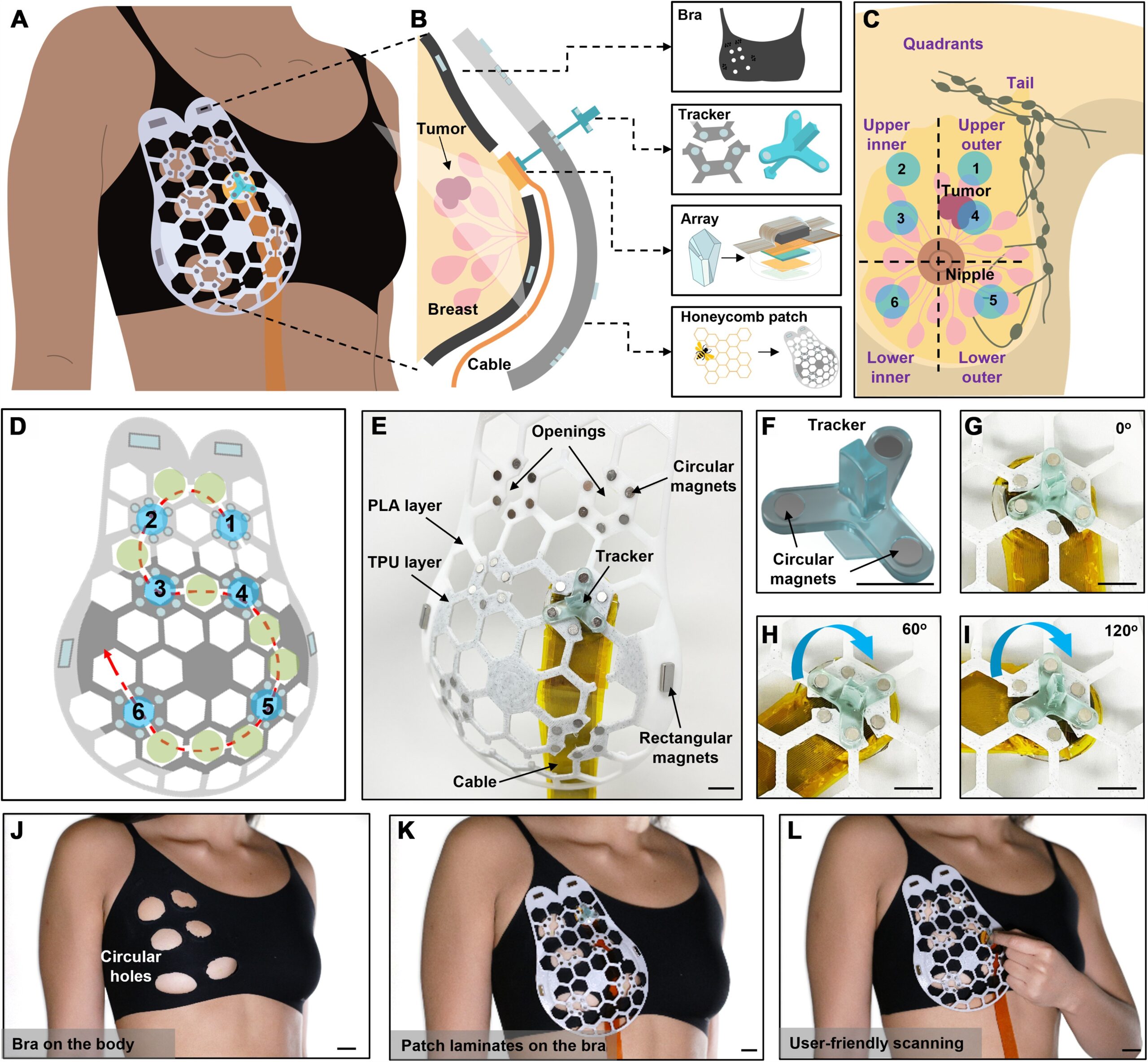
Follow this link to see a detailed caption for this graphic (labeled Fig. 1E) in the published study.
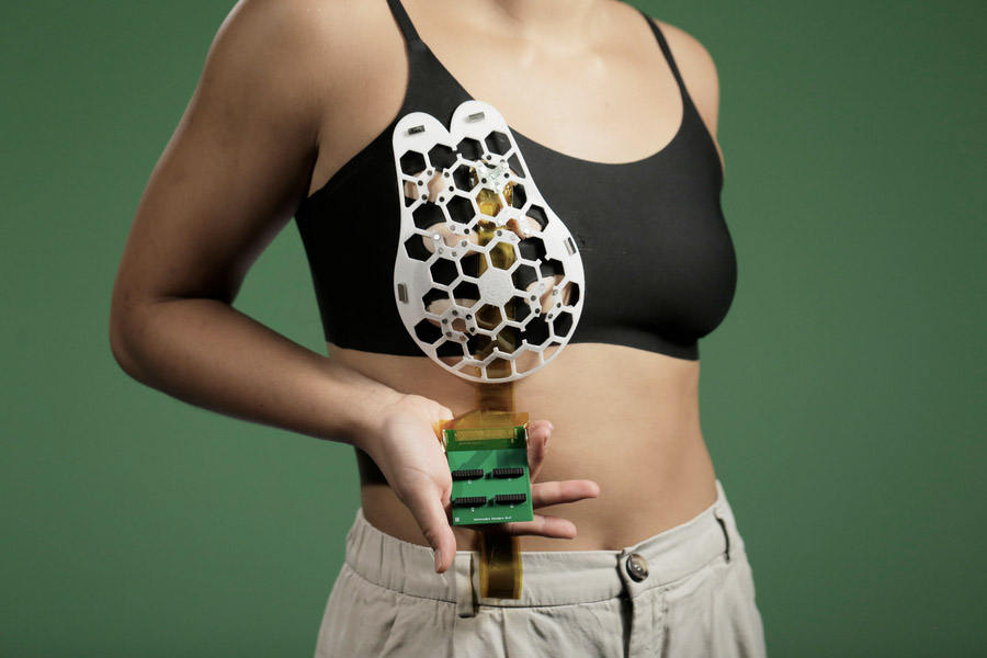
The Conformable Ultrasound Breast Patch, dubbed the cUSBr-Patch, is a flexible patch with honeycomb-like openings that can be attached to a bra using multiple tiny magnets, allowing the wearer to move an ultrasound tracker along the patch to six different positions and image the breast tissue from different angles. In the new study, according to MIT News, the researchers showed that they could obtain ultrasound images with a resolution comparable to that of the ultrasound probes used in medical imaging centers.
“We changed the form factor of the ultrasound technology so that it can be used in
your home,” says Dagdeviren. “It’s portable and easy to use, and provides real-time, user-friendly monitoring of breast tissue.” The wearable ultrasound patch can be used over and over and does not require any special expertise to operate. This video demonstrates how it works.
The MIT team created the nature-inspired honeycomb design using Autodesk Fusion 360 software and designed it with four main criteria in mind: 1) ease of use, 2) range of motion, requiring 360-degree rotation at a given position with a repeatable array localization), 3) sustainable manufacturing, and 4) user comfort.
Honeycomb structures are often preferred in medical devices because they minimize the amount of material used in manufacturing, maximize flexibility, easily cover large areas of the skin, and provide structural stability, allowing for the conformability of the patch and the customization of array scanning.
3D printing with TPU and PLA
The researchers used a basic 3D printer to produce a prototype, a 2 mm-thick patch with two different layers of material –– a white thermoplastic polyurethane (TPU) layer and a gray-colored polylactic acid (PLA) layer, achieving a dual effect of conformability to the body and strong rigidity in the structure of the patch. They started the print with a white TPU filament at 100 percent infill and programmed it to pause at a specific layer of the patch. They then exchanged the TPU for PLA and continued until the part was completed.
The researchers used an Elegoo Mars 2 resin printer to print the tracker with an ABS-like photopolymer resin material. They then cured it for strength using a UV Cure Box machine to ensure that the tracker was strong enough to withstand the torsion forces experienced during the carrying and rotating of the one-dimensional (iD) phased array.
Anantha Chandrakasan, dean of MIT’s School of Engineering and one of the authors of the study, stated: “This work will significantly advance ultrasound research and medical device designs, leveraging advances in materials, low-power circuits, AI algorithms, and biomedical systems.”